
TFAST®とは?
TFAST®(ティファーストもしくはティファスト)とはアメリカで開発された胸部の迅速超音波検査法です。胸の中に胸水や心膜浸出液などがあるか? 心臓や肺に異常はないかを検査します。以前は交通事故や落下時などに救急時に用いられた検査法ですが、現在ではより進化して、身体検査や聴診についで行われるスクリーニング検査と言っても過言でなくなっています。TFAST®では今まで見過ごされていた多くの病気や体の状態を迅速に把握することができ、海外ではすべての獣医師が身につけるべき、次世代のコア・スキルと言われています。TFAST®︎はGlobalFAST®︎の一部でもあります。
当院では、院長が長年取締役を務める動物病院向け情報配信会社「ペット・ベット社」(VMN:Veterinary Medical Networkを運営)によるプロジェクトの一環として、TFAST® のオンラインセミナーを翻訳し、日本の動物病院への紹介・普及に努めています。
獣医師の先生方には、「FastVET.com オンラインセミナー日本語版」
• [AFAST®︎(腹部)迅速超音波検査法]
をぜひ受講いただき、グローバルスタンダードな獣医超音波診断スキル を身につけてください。
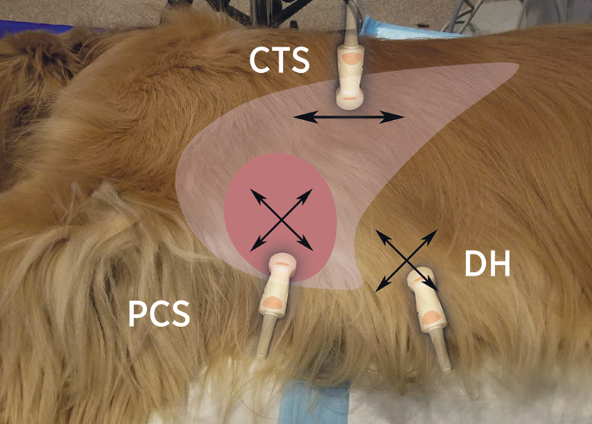
検査方法
超音波診断装置により、胸部を観察します。
TFAST® 検査よってできること
- 胸水(遊離体液)の識別:検出率はレントゲン検査よりも優れています。
- 心膜浸出液(貯留液)の検出 – FASTVetTM NEW
- *気胸 (遊離した気体)の検出 – FASTVetTM NEW
- 血液量の状態 & 収縮性 左室短軸像から – FASTVetTM NEW
- 長軸像での右室対左室比 (RV:LV)右心の病態
- 短軸像での左房対大動脈比 (LA:Ao) 左心の病態
- 後大静脈と肝静脈の分類による血液量の状態の評価 – FASTVetTM NEW
- 目的臓器アプローチによる心臓と胸部の軟部組織異常の検出
- TFAST CTS 画像からの延長でVet BLUE® に移ることができる– FASTVetTM NEW
access_time
検査時間
- 検 査:TFAST®のみだと5〜10分程度程かかります。
- 検査結果:最終報告書作成までは1時間以上かかることがありますので、時には検査結果だけお伝えして、報告書は後日お渡しとなることもあります。
warning
注意事項
- TFAST®では基本的に検査に先立ち、剃毛(毛刈り)は通常行いませんので、ご安心ください。
※AFAST®、TFAST®、VetBlue®、GloubalFAST®という用語は、その完全な明確性と高品質な教育目的事業を維持するため、米国特許商標局に登録されており、Lisciandro Enterprises PLLCが所有しています。また、すべてのFASTVetの資料は著作権により保護されていますので、無断複写・転載は禁止されています。VMN:Veterinary Medical Networkを運営する(株)ペット・ベット社では契約により許可を得てこれらを正規に使用しています。
library_books
参考文献・資料等
- Point-of-Care Ultrasound in Medical Education — Stop Listening and Look
- Evaluation of a thoracic focused assessment with sonography for trauma (TFAST) protocol to detect pneumothorax and concurrent thoracic injury in 145 traumatized dogs
- The use of the diaphragmatico-hepatic (DH) views of the abdominal and thoracic focused assessment with sonography for triage (AFAST/TFAST) examinations for the detection of pericardial effusion in 24 dogs (2011-2012)
- Thirteen dogs and a cat with ultrasonographically detected gallbladder wall edema associated with cardiac disease
- TFAST Accurate Diagnosis of Pleural and Pericardial Effusion, Caudal Vena Cava in Dogs and Cats
- Abdominal and thoracic focused assessment with sonography for trauma, triage, and monitoring in small animals
- POCUS: TFAST-Introduction and Image Acquisition
- Cageside Ultrasonography in the Emergency Room and Intensive Care Unit.
- Focused ultrasound of the caudal vena cava in dogs with cavitary effusions or congestive heart failure: A prospective, observational study
- Thirteen dogs and a cat with ultrasonographically detected gallbladder wall edema associated with cardiac disease
- TFAST Accurate Diagnosis of Pleural and Pericardial Effusion in Dogs and Cats.
- A Whole-Body Approach to Point of Care Ultrasound
![]()
・
![]()
<1>